Make 2015 the Year of the Smile with Teeth Whitening from Horsham Dental Elements

January 20, 2015

100047604The New Year has really just gotten started, so it’s not too late to make one more resolution. Resolve to smile more! Smiling isn’t just good for you, it’s good for everyone around you, too—acquaintances and strangers alike. And if you really want your smile to have an impact, then consider teeth whitening with Dr. Gary Henkel of Horsham Dental Elements in Horsham, PA, and Philadelphia, PA.

Teeth can become dull and discolored for a number of reasons, including:


  • The natural aging process
  • Favorite foods and beverages, such as coffee, tea, cola, blueberries, cherries and chocolate
  • Certain prescription medications, such as the antibiotic tetracycline
  • Teeth grinding, which may cause the biting edge of teeth to darken
  • Tooth injury that leads to cracks, which hide stain-causing debris

Teeth whitening doesn’t just improve the appearance of your smile. Numerous studies have shown that people with white teeth are more confident, more socially out-going and are even considered to be smarter than people whose smiles aren’t as white. So, a white smile not only goes a long way aesthetically, it can impact you emotionally and psychologically.

Teeth Whitening Made Easy

Dr. Henkel and his staff at Horsham Dental Elements understand that your schedule is busy enough already. You shouldn’t have to squeeze another appointment onto your calendar to have white teeth. That’s why we offer an at-home teeth whitening kit for you to use in the privacy and convenience of home. We’ll give you custom-fit whitening trays that you’ll fill with the professional grade gel we supply. Then, just wear the trays as Dr. Henkel instructs, and you’ll be on your way to a whiter and brighter smile in no time!

Call Our Office Today

If you would like to learn more about at-home teeth whitening, then call Dr. Henkel at Horsham Dental Elements in Philadelphia, PA, and Horsham, PA. We also serve patients from Dresher, Springfield and Hatboro, as well as Bucks County and Montgomery County.

Cosmetic Bonding from Dr. Henkel Solves Minor Tooth Flaws

November 15, 2014

158554263Accidents happen, well, accidentally. Unfortunately, our teeth are sometimes hurt in accidents. Like when you bite down on a hard candy and chip a tooth. On the other hand, accidents aren’t always the culprit of minor tooth imperfections. Perhaps you have a few teeth that are misshapen or slightly misaligned. In either case, cosmetic bonding may be the answer. Dr. Gary Henkel at Horsham Dental Elements in Philadelphia, PA, and Horsham, PA, has repaired the smiles of many patients thanks to cosmetic bonding.

Cosmetic Bonding is a common dental procedure where a tooth-colored plastic dental material is applied to the tooth and hardened with a special light. The bonded material restores the tooth’s natural appearance and functionality. Cosmetic bonding is often the best option for when:

  • A tooth is chipped or cracked
  • A tooth requires a filling because of decay
  • Teeth are discolored
  • Teeth are misaligned or have gaps in between them
  • A tooth is misshapen or should look longer

Cosmetic Bonding: As Easy as 1-2-3

As dental procedures go, cosmetic bonding is relatively simple. Dr. Henkel will use a shade chart to make sure the bonding material is a match to your tooth color. Then, the tooth’s surface is made slightly rough, which allows the bonding material to adhere more securely to the tooth. After Dr. Henkel applies the resin, he will mold and smooth it to the desired shape. Once the special light hardens the material, the doctor may need to further shape the bonding and polish it to match the existing tooth. In less than an hour, you’ll have a tooth that looks as good as new!

Call Our Office Today

If you’ve been contending with a flawed tooth for a while—or if an accident just happened—then schedule an appointment for cosmetic bonding with Dr. Henkel today. Horsham Dental Elements in Philadelphia, PA, and Horsham, PA, also serves patients in Dresher, Springfield and Hatboro as well as Bucks County and Montgomery County.

Get to the Root of the Problem with Root Canal Therapy

October 14, 2014

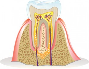
480377293Sometimes, a root canal is the retort to an offer of something you really don’t want—as in, “I want that like I want a root canal.” But if you are experiencing the pain of severe tooth decay or an infected tooth, then the best choice may indeed be a root canal. Dr. Gary Henkel at Horsham Dental Elements in Philadelphia, PA, and Horsham, PA, is an expert in root canal therapy and uses state-of-the-art equipment to ensure that you receive the finest care possible.

What is Root Canal Therapy?

A root canal is the terminology used to describe the procedure used to save a badly decaying or infected tooth. During a root canal, Dr. Henkel—who lectures on endodontics and provides consulting services to three endodontic supply manufacturers or suppliers—removes the nerve, blood and soft tissue inside the canal. Then, the canal is thoroughly cleaned of decay and debris, and finally Dr. Henkel fills the canal to prevent further decay. Throughout the procedure, Dr. Henkel uses a surgical microscope for magnification and the best visibility possible during your root canal.

When is Root Canal Therapy Necessary?

Well first of all, if you are experiencing the pain of a severely decayed or infected tooth, a root canal will eliminate that pain. Secondly, the bacteria that causes the decay and infection can spread beyond the root of the tooth, bringing about an abscess. Although there are instances when no symptoms are present, some of the signs that may signal the need for a root canal include:

  • Extreme tooth pain when chewing
  • Sensitivity to hot or cold
  • A darkening color of the tooth
  • Swelling of the gums around the tooth

With proper general dentistry care, a root canal may be avoidable. If one is necessary, however, rapid attention is required to head off further trouble, such as gum disease or the spread of bacteria throughout the body.

Call Our Office Today

If you suspect that you may need root canal therapy, schedule an appoint today with Dr. Gary Henkel at Horsham Dental Elements in Philadelphia, PA, or Horsham, PA. Our offices also serve patients in surrounding Bucks county and Montgomery county, as well as Hatboro, Springfield and Dresher.

Revitalize the Beauty of your Smile with Cosmetic Options at Horsham Dental Elements

July 1, 2013

While Dr. Henkel and his dedicated dental staff believe strongly that optimal oral health should come first, they also understand that many patients are interested in having a more beautiful smile as well. After all, it’s not hard to understand why – white, straight teeth can imply all sorts of positive attributes about you to your peers and coworkers, including confidence, attractiveness, youth, vitality, and much more. Our team at Horsham Dental Elements in Horsham and Philadelphia would be more than happy to help you achieve this dream with the help of multiple cosmetic dentistry options designed to rejuvenate smiles and help them shine again! We offer:

  • Both in-office and take-home teeth whitening to accommodate busy schedules
  • Cosmetic bonding to reshape broken or worn-down teeth
  • Porcelain veneers to cover your teeth and give them a straighter, whiter, and healthier appearance
  • Invisalign to quickly and seamlessly realign crooked teeth

Additionally, several of our restorative dentistry procedures have valuable cosmetic benefits, such as dental implants, which seamlessly fill unsightly gaps in your smile with beautiful, durable replacements, porcelain crowns, and tooth-colored fillings. Rest assured that we’ll take every precaution to ensure that any work you receive at our offices is both durable and natural-looking, giving you a look that you can feel proud of for years to come!

Contact either our Horsham or Philadelphia office if you’d like to learn more about we can turn your smile into a stunner, or if you’re ready to go ahead and schedule an appointment. Dr. Henkel is well-known for his thorough, compassionate care that delivers excellent results and sends patients off with a new reason to smile. We’re also happy to serve the areas of Montgomery, Buck County, and beyond.

BISPHOSPHONATE COATED IMPLANTS?????

March 14, 2012

Filed under: Uncategorized — admin @ 10:32 am

Strange as it may sound, implants with bisphosphonate coatings have entered human trials, with reported stronger connections of bone to the implant. here is a summary of the research:


New method for stronger dental implants

Millions of people have bad teeth replaced with implants. Often following the procedure, they are unable to chew food for up to six months, until the implant has become fixated in the bone. Now, for the first time, a drug coating that has been tested on humans allows titanium screws to adhere to the bone better and faster. The Linköping researchers behind the method report that the results are good.
En käke med två olika implantat
The study, led by Per Aspenberg, professor of orthopaedic surgery at Linköping University, is published in the journal Bone and was highlighted in this week’s edition of the British Medical Journal (BMJ).
The implants are screwed into the jawboneand provide purchase for artificial teeth. Using current technology, it may take four to six months before the bone surrounding the screw has healed and is strong enough so the patient can begin to benefit from surgery.
The coating, developed at Linköping University in Sweden, consists of a nanometre-thin layer of protein that attaches to the metal surface. Attached to the protein is a drug belonging to the bisphosphonates, usually used to treat osteoporosis. Several animal studies have shown that this method allows the bone surrounding the implant to rapidly become denser and stronger.
Now, for the first time, this method has been tested on humans. 16 patients each received two implants; one normal and a similar surface-treated implant as described above. Neither the patient nor the dental surgeon knew which was which. After six months it was noted that for 15 of the 16 patients the treated screw was markedly much better established. Already after two months X-ray images showed positive changes adjacent to the treated screws. No complications occurred.
“It is the first time ever anyone has succeeded in reinforcing the bone around an implant with localised medication”, says Per Aspenberg, professor of orthopaedics, who devised the method of using bisphosphonates in this way.
Pentti Tengvall, professor of biomaterials, developed the method of adhering the drug to the screw and the study was conducted by Jahan Abtahi, specialist MD and PhD, supported by Per Aspenberg. Addbio AB is a Linköping based company now working on commercializing the surface treatment for different types of implants in bone.
Article:A bisphosphonate-coating improves the fixation of metal implants in human bone. A randomized trial of dental implants by Jahan Abtahi, Pentti Tengvall and Per Aspenberg. The journal ‘Bone’ (in press), published online 10 February 2012.
Contact:Per Aspenberg +46 733 866 468
Related Links
osteoklasterna anfaller
Osteoclasts are giant bone-eating cells and when they attack, the bone is broken up and the implant loosens. The new coating is resistant to attack.
Drawing: Per Aspenberg


Åke Hjelm 2012-02-29


Harvesting autogenous bone made quick and easy

March 5, 2012

Filed under: Uncategorized — admin @ 11:50 am

Recently, Neo Biotech in Korea has developed a special trephine bur with a sleeve they call the ACM or Auto Chip Maker.  This unique device fits in the standard osteotomy latch type handpiece, is run between 300 to 500 rpms, and will penetrate 4mm deep to a safety stop, each penetration yielding about one cc of autogenous particulate.  I have posted an eight minute video showing it being used to repair an ailing implant on you tube, which can be seen here:

http://www.youtube.com/watch?v=GqICDDfXM-A&list;=UUU49GWbwWG0uiwmbhm1xnyw&index;=1&feature;=plcp

Periapical pathology may not be a contraindication for immediate implant placement

February 1, 2012

Filed under: Uncategorized — admin @ 10:39 am
Background: Many patients requiring implant therapy present with hopeless teeth exhibiting periapical pathology. The
advisability of implant placement in such situations has not
been conclusively determined.
Methods: Sixty-four patients underwent therapy in their
maxillary incisor region. Treatment consisted of immediate
implant placement in a site demonstrating periapical pathology, and immediate implant placement in a ‘‘pristine’’ site,
either during the same visit or during separate visits. The implants placed in the sites demonstrating periapical pathology
were followed in function for £117 months, with a mean time in
function of 64 months. The implants placed in pristine sites
were followed in function for £120 months, with a mean time
in function of 62 months.
Results: Two implants in the central incisor positions of one
patient demonstrated 2 mm of buccal recession after 46
months in function. These implants were deemed esthetic failures, despite the absence of in?ammation and continued clinical implant immobility, yielding cumulative survival rates of
98.1 and 98.2 for implants placed in sites with periapical pathology and implants placed in sites without periapical pathology, respectively, according to published criteria.
Conclusions: Implants immediately placed in sites demonstrating periapical pathology yielded results comparable to
those immediately placed in pristine sites. The difference in
survival rates was not statistically signi?cant. J Periodontol
2012;83:182-186.
KEY WORDS
Dental implants; dental materials; pathology, oral; peri

PERI IMPLANTITIS MORE PREVALENT IN SMOKERS AND THOSE WITH PERIO DISEASE

November 3, 2011

Filed under: Uncategorized — admin @ 10:22 am

A recent publication out of sweden showed a positive correlation between peri implantitis and previous perio disease and smoking.  the abstract is post here:

Swed Dent J. 2010;34(2):53-61.

Peri-implantitis in a specialist clinic of periodontology. Clinicalfeatures and risk indicators.

Source

Department of Periodontology at Kista-Skanstull, Folktandvården i StockholmsIän AB, Stockholm, Sweden.

Abstract

Implant therapy has become a widely recognizedtreatment alternative for replacing missing teeth. Several long term follow-upstudies have shown that the survival rate is high. However, complications mayappear and risk indcators associated with early and late failures have beenidentified. The purpose of the present retrospective clinical study was todescribe some clinical features of patients with clinical signs ofperi-implantitis and to identify risk indicators of peri-implantitis in apopulation at a specialist clinic of Periodontology. In total,the materialconsisted of 377 implants in 111 patients with thediagnosis peri-implantitis. The mean age at the examination was found to be56.3 years (range 22-83) for females and 64.1 years (range 27-85) for males.The mean number of remaining teeth was found to be 10.5 (S.D. 8.89) and themean number of implants was 5.85 (S.D. 3.42). Fora majority of the subjects, more than 50% of the remaining teeth had a marginalbone loss of more than 1/3 of the root length. Forty-sex percent of thepatients visited regularly dental hygienists for supportive treatment. Thepercentage of implants with peri-implantitis wassignificantly increased for smokers compared to non-smokers (p = 0.04). In thegroup of non-smokers, 64% of the implants had thediagnosis peri-implantitis, while the corresponding relative frequency forsmokers was 78%. A majority of the individuals had a Plaque index and Bleedingon probing index >50%. The median of the follow-up time after implantplacement was 7.4 years and the observation period was not significantlycorrelated to the degree of bone loss around the implants.Among the subjects with a mean bone loss >6 mm at implantswith peri-implantitis, more than 70% had a mean marginal bone loss > 1/3 ofthe root length of the remaining teeth. A positive and significant correlationwas found between the degree of marginal bone loss in remaining teeth and thedegree of bone loss around implants withperi-implantitis. In conclusion, the results of the present study indicate thatsmoking as well as previous history of periodontitis are associated withperi-implantitis and may represent risk factors for this disease


MIX ACETOMINOPHEN AND IBUPROFEN FOR BEST RESULTS

November 1, 2011

Filed under: Uncategorized — admin @ 3:10 pm


Dr. Richard L. Wynn is Professor of Pharmacology at the University of Maryland Dental School. He holds a BS degree in Pharmacy and a PhD degree in Pharmacology. He chaired the Department of Pharmacology at the University of Maryland Dental School from 1980 to 1995. He is the lead author of the Drug Information Handbook for Dentistry, a co-author on many other dental drug publications, an author of over 300 refereed scientific journal articles, a consultant to the Academy of General Dentistry, a featured columnist, and a featured speaker presenting more than 500 courses in continuing dental education. One of his primary interests continues to be keeping dental professionals informed of all aspects of drug use in dental practice.

Over-the-Counter Ibuprofen and Acetaminophen in Combination are Superior to Each Agent Alone in Treating Postoperative Pain

The first two reports (Derry, et al and Toms, et al) describe the efficacy of single doses of ibuprofen and single doses of acetaminophen for postoperative pain, compared to placebo in well-controlled trials. The third report (Mehlisch, et al) describes the analgesic benefits of the combination of the two agents (compared with each drug alone), again in well-controlled trials. These studies provide further evidence-based information for the rational use of over-the-counter pain relievers for postoperative dental pain.
Study #1 Assessing the efficacy of single dose over-the-counter ibuprofen
Derry C, Derry S, Moore RA, et al, “Single Dose Oral Ibuprofen for Acute Postoperative Pain in Adults,” Cochrane Database Syst Rev, 2009, July 8, (3) CD001548.
The objective of this study was to assess the analgesic efficacy of ibuprofen in single oral doses for moderate and severe postoperative pain in adults. The method was to search the Cochrane Library, CENTRAL, MEDLINE, EMBASE, and the Oxford Pain Relief Database for studies to May, 2009. Selection of studies considered for review was based on randomized, double-blind, placebo-controlled trials of single-dose orally administered ibuprofen in adults with moderate to severe acute postoperative pain. Pain relief or pain intensity data were extracted and converted to outcomes of numbers of participants with at least 50% pain relief over 4 to 6 hours, from which relative risk and number-needed-to-treat-to-benefit (NNT) were calculated. The number of participants using rescue medication over specified time periods, and the time to rescue medication, were sought as additional measures of efficacy. Finally, information on adverse events and withdrawals was collected.
Results of the Derry, et al, study
The studies reviewed totaled 72 and included 9,186 participants, in which ibuprofen was compared to placebo. The majority of the studies used ibuprofen at 200 mg and 400 mg doses.
For at least 50% pain relief compared with placebo, the number-needed-to-treat-to-benefit for ibuprofen 200 mg (2,690 participants) was 2.7 (2.5 to 3.0) and for ibuprofen 400 mg (6,475 participants) it was 2.6 (2.4 to 2.6).
The proportion with at least 50% pain relief was 46% with 200 mg ibuprofen and 54% with 400 mg ibuprofen. Remedication within 6 hours was less frequent with higher doses, with 48% remedicating with 200 mg and 42% remedicating with 400 mg. The median time to remedication was 4.7 hours for 200 mg and 5.4 hours for 400 mg.
The studies using dental impaction pain models and soluble ibuprofen salts (liquid gel capsule formulations) produced better efficacy estimates. Adverse events were uncommon and no different from placebo.
The authors concluded a substantial amount of high quality evidence demonstrated that over-the-counter ibuprofen is an effective pain reliever in treating postoperative pain.
Study #2 Assessing the efficacy of single dose over-the-counter acetaminophen
Toms L, McQuay HJ, Derry S, et al, “Single Dose Oral Paracetamol (Acetaminophen) for Postoperative Pain in Adults,” Cochrane Database Syst Rev, 2008, Oct 8, (4) CD004602.
The objective of this study was to assess the efficacy of single dose oral acetaminophen for the treatment of acute postoperative pain. The method was to search the Cochrane Library, MEDLINE, EMBASE, the Oxford Pain Relief Database, and reference lists of articles to update data from an existing version of the review. Selection of studies considered for review was based on randomized, double-blind, placebo-controlled trials of acetaminophen for acute postoperative pain in adults.
Area under the “pain relief versus time” curve was used to derive the proportion of participants with acetaminophen or placebo experiencing at least 50% pain relief over 4 to 6 hours, using validated equations. Number-needed-to-treat-to-benefit was calculated. The proportion of participants using rescue analgesia over a specified time period and time to use were sought as measures of duration of analgesia. Information on adverse events and withdrawals was also collected.
Results of the Toms, et al, study
Fifty-one studies with 5,762 participants were included; of those who took acetaminophen or placebo, 3,277 participants were treated with a single oral dose of acetaminophen and 2,425 with placebo. About half of the participants treated with acetaminophen at standard doses achieved at least 50% pain relief over 4 to 6 hours compared with about 20% treated with placebo.
Number-needed-to-treat-to-benefit for at least 50% pain relief over 4 to 6 hours following a single 500 mg dose of acetaminophen was 3.5 (2.7 to 4.8); following a single 600 to 650 mg dose it was 4.6 (3.9 to5.5); following a single dose of 975 to 1000 mg it was 3.6 ( 3.4 to 4.0).
About half the participants taking acetaminophen needed additional analgesia over 4 to 6 hours compared with about 70% with placebo.
Five people would need to be treated with 1000 mg acetaminophen, the most commonly used dose, to prevent one needing rescue medication over 4 to 6 hours, who would have needed it with placebo.
Reported adverse events were mild and transient, and occurred at similar rates with 1000 mg acetaminophen and placebo. No serious adverse events were reported.
The authors concluded that a single dose of acetaminophen provided effective analgesia for about half of the patients with acute postoperative pain for a period of about 4 hours and is associated with few, mainly mild, adverse events.
Study #3 Assessing the efficacy of the combination of over-the-counter ibuprofen and acetaminophen
Mehlisch DR, Aspley S, Daniels SE, et al, “Comparison of the Analgesic Efficacy of Concurrent Ibuprofen and Paracetamol With Ibuprofen or Paracetamol Alone in the Management of Moderate to Severe Acute Postoperative Dental Pain in Adolescents and Adults: A Randomized, Double-blind, Placebo-controlled, Parallel-group, Single-dose, Two-center, Modified Factorial Study,” Clin Ther, 2010, 32:882-95.
This study was done to test the hypothesis that combination analgesics may offer improved analgesic efficacy, particularly for moderate to severe pain. The authors evaluated the analgesic benefits of concurrent ibuprofen and paracetamol (acetaminophen), compared with each drug used alone, in the management of acute postoperative dental pain.
Methods
The study enrolled healthy patients, ages 16 to 40 years, undergoing surgical removal of 3 to 4 impacted molars. The protocol was a randomized, double-blind, placebo-controlled, parallel-group, single-dose, two center study. Patients were randomly assigned to ibuprofen 400 mg/acetaminophen 1000 mg combination, ibuprofen 200 mg/acetaminophen 500 mg combination, ibuprofen 400 mg alone, acetaminophen 1000 mg alone, or placebo when the postoperative pain reached moderate to severe intensity.
The primary endpoint of efficacy was the sum of pain relief and pain intensity differences from 0 to 8 hours. Secondary endpoints included total pain relief, sum of pain intensity differences, and sum of pain intensity differences on the visual analog scale at various time endpoints. Other analgesic measures included peak effect, onset and duration of effect, and patients’ overall assessment of treatment. The tolerability of study medicines was also assessed in terms of frequency and nature of adverse events.
Results of the Mehlisch, et al, study
A total of 234 patients were randomly assigned to treatment and included in the intent-to-treat population.
Results for the primary endpoint
For the sum of pain relief and pain intensity differences, the group receiving the combination of ibuprofen 400 mg/acetaminophen 1000 mg had significantly better mean scores compared with ibuprofen 400 mg alone, acetaminophen 1000 mg alone and the combination of ibuprofen 200 mg/acetaminophen 500 mg.
For the sum of pain relief and pain intensity differences, the group receiving the combination of ibuprofen 200 mg/acetaminophen 500 mg had significantly better mean scores compared with acetaminophen 1000 mg alone, but not compared to ibuprofen 400 mg alone.
Results for secondary endpoints
Ibuprofen 400 mg/acetaminophen 1000 mg was associated with significantly better scores than was single agent therapy for total pain relief, sum of pain intensity differences, and sum of pain intensity differences on the visual analog scale at all time intervals, and for sum of pain relief and pain intensity differences from 4 to 6 hours.
A breakdown of the Mehlisch, et al, data showed the following:
Time to the first confirmed perceptible pain relief for each treatment was:
Ibuprofen 400 mg/acetaminophen 1000 mg: 23 minutes
Ibuprofen 200 mg/acetaminophen 500 mg: 22 minutes
Ibuprofen 400 mg: 49 minutes
Acetaminophen 1000 mg: 25 minutes
Placebo: 98 minutes
Time to the first meaningful pain relief for each treatment was:
Ibuprofen 400 mg/acetaminophen 1000 mg: 94 minutes
Ibuprofen 200 mg/acetaminophen 500 mg: 74 minutes
Ibuprofen 400 mg: 124 minutes
Acetaminophen 1000 mg: 141 minutes
Placebo: 133 minutes
Time to pain half gone for each treatment was:
Ibuprofen 400 mg/acetaminophen 1000 mg: 70 minutes
Ibuprofen 200 mg/acetaminophen 500 mg: 86 minutes
Ibuprofen 400 mg: 114 minutes
Acetaminophen 1000 mg: 135 minutes
Placebo: 229 minutes
There were significant differences in favor of all active treatments versus placebo for all efficacy endpoints. Adverse events were similar across treatments, with the most frequent being nausea [26.1% (61/234)], vomiting [18.8% (44/234)], headache [10.3% (24/234)], and dizziness [8.1% (19/234)].
The authors concluded that concurrent ibuprofen and acetaminophen appeared to provide significantly better analgesic efficacy compared with either drug alone for management of acute postoperative dental pain.


Drug information is constantly changing. Promote medication safety in your practice with Lexicomp Online for Dentistry.

PERI-IMPLANTITIS AND ITS MANAGEMENT

October 26, 2011

Filed under: Uncategorized — admin @ 1:10 pm

Review of the treatment protocols for peri-implantitis

Oct 12, 2011

By Scott Froum, DDS

Peri-implantitis is an inflammatory process affecting the soft and hard tissues surrounding an implant. This disease is associated with loss of supporting bone, bleeding on probing, and occasionally suppuration. The etiopathogenesis of peri-implantitis is complex and related to a variety of factors that affect the peri-implant environment. Peri-implantitis can be influenced by three factors:

  1. Patient-related factors including systemic diseases (e.g, diabetes, osteoporosis) and prior dental history (periodontitis)
  2. Social factors such as inadequate oral hygiene, smoking, and drug abuse
  3. Parafunctional habits (bruxism and malocclusion).

In addition to the above, iatrogenic factors such as faulty restorations, cement left following restoration delivery, and/or loose components can also play a significant role in the development of peri-implantitis.

Although restorations of endosseous implants have demonstrated a very high survival rate1, one study suggested that over a five-year period, 0 to 14.4% of dental implants demonstrated peri-implant inflammatory reactions associated with crestal bone loss.2

Many methods of treating peri-implantitis have been documented in the literature and most focus on removal of the contaminating agent from the implant surface. These treatments include:

  1. Administration of systemic antibiotics alone
  2. Mechanical debridement with or without systemic antibiotic treatment3
  3. Mechanical debridement with or without localized drug delivery and chlorhexidine oral rinses3a
  4. Mechanical debridement combined with LASER decontamination4
  5. Surgical debridement, and more recently …
  6. Surgical debridement with guided bone regeneration (GBR) for reparation of bony and soft-tissue defects.5

To date, GBR using a bone graft and membrane has had the best success as in demonstrating bone fill of the defects associated with peri-implantitis as described in the literature.6

Because there are biologic differences between teeth and implants, the progression of infection around implants is different than natural teeth. The inflammatory cell infiltrate around implants was reported to be larger and extend more apically when compared to a corresponding lesion in the gingival tissue around natural teeth. In addition, the tissues around implants are more susceptible to plaque-associated infections that spread into the alveolar bone.7

Implant surface bacterial decontamination is essential in treating peri-implantitis infections. Systemic administration of antibiotics has been used in the treatment of peri-implantitis, resulting in a reduction of inflammation. However, the efficacy of antibiotic therapy as a sole therapy has limited efficacy due to bacterial recolonization of the implant surface.8

Because nonsurgical treatment approaches failed to promote the reosseointegration of the exposed implant sites,9additional surgical interventions have been used in order to minimize the risk for a reinfection of the peri-implant pocket. Some of the treatment modalities suggested for peri-implantitis are:

  1. Mechanical/ultrasonic debridement with localized drug delivery; i.e., antimicrobial minocycline spheres (Arestin®)
  2. Laser treatment with and without flap access
  3. Open flap debridement
  4. Open flap debridement with guided bone regeneration

Renvert et al. (2006) compared the combination of mechanical debridement and topical application of minocycline microspheres to the combination of mechanical debridement and 1% chlorhexidine gel application in peri-implant lesions. The results obtained after a follow-up period of 12 months showed that with the chlorhexidine group, only a limited reduction in bleeding on probing was achieved and the mean peri-implant probing depth (PD) remained unchanged (3.9 mm). On the other hand, in the minocycline group, the reduction of bleeding on probing was greater and an improvement in mean peri-implant PD (from 3.9 mm to 3.6 mm) was seen. These results suggest that the topical application of chlorhexidine provides limited or no adjunctive clinical improvement when treating shallow peri-implant lesions as compared with using mechanical debridement alone.10

Schwarz et al. (2005, 2006a) compared the efficacy of the Er:YAG laser with that of the combination of mechanical debridement (using plastic curettes) and antiseptic (0.2% chlorhexidine digluconate) administration for the treatment of peri-implantitis. In both studies, the results obtained six months after therapy suggested that the treatment modalities were equally effective in improving peri-implant probing pocket depth (PPD) and clinical attachment level (CAL). However, at 12 months, the mean values for both groups of peri-implant PPD and CAL were not statistically significantly different from the corresponding values at baseline. Therefore, the results of the Er:YAG laser seem to be limited to a six-month period, particularly for advanced peri-implantitis lesions.

Leonhardt et al. evaluated the five-year outcome of a combined surgical (open flap) and antimicrobial treatment of peri-implantitis in humans. Leonhardt studied the effect of systemic antimicrobial therapy (amoxicillin and metronidazole) together with an open flap procedure and in conjunction with mechanical debridement of the implant surface for decontamination. The treatment was successful in 58% of the implants treated during follow-up for five years. Smoking was found to be a negative risk factor for treatment success.11

Schwarz et al. (2006b) evaluated and compared the efficacy of two bone regenerative procedures for the treatment of moderate intrabony peri-implantitis lesions that included a greater than 6 mm probing depth and an intrabony component of 3 mm as detected on radiographs. The defects were randomly treated either with a surgical debridement and filled with nanocrystalline hydroxyapatite, or surgical debridement and filled with bovine-derived xenograft (Bio-Oss®, Geistlich, Wolhusen, Switzerland) combined with a bioresorbable porcine-derived collagen membrane (Bio-Gide®, Geistlich, Wolhusen, Switzerland). After two years, the study showed that the combination of bovine bone mineral and the collagen membrane seemed to yield greater improvements in clinical parameters, showing 0.9 ± 0.2 mm more in PD reduction and 1.0 ± 0.3 more clinical attachment gain.12,13

Conclusion
The ideal management of peri-implant infections should focus both on infection control of the lesion, detoxification of the implant surface, and regeneration of lost support. Treatment options can be surgical or nonsurgical. To date, studies suggest that nonsurgical treatment of peri-implantitis is unpredictable, and the use of chemical agents such as chlorhexidine has only limited effects on clinical and microbiological parameters. Adjunctive local or systemic antibiotics have shown to reduce bleeding on probing and probing depths in combination with mechanical debridement. Beneficial effects of laser therapy on peri-implantitis have been shown, but this approach needs to be further evaluated. Implant surface bacterial decontamination is essential in treating peri-implantitis infections. Most studies suggest that establishing an adequate healthy peri-implant tissue environment proves to be difficult since inflammation is still present in a significant number of patients. New treatment modalities need to be evaluated using long-term randomized-controlled studies to identify predictable and successful treatment of peri-implantitis.

Author bio
Scott Froum, DDS, is a periodontist and co-editor of Surgical-Restorative Resource e-newsletter. He is a clinical associate professor at the New York University Dental School in the Department of Periodontology and Implantology. He is in private practice in New York City. You may contact him by email at drscottfroum@yahoo.com.

References
1. Albrektsson T et al. Osseointegrated oral implants — A Swedish multicenter study of 8139 consecutively inserted Nobelpharma implants. J Periodontol 1988;5 9:287-296.
2. Berglundh T et al. A systematic review of the incidence of biological and technical complications in implant dentistry reported in prospective longitudinal studies of at least 5 years. J Clin Periodontol 2002; 29:197-212.
3. Renvert S. (2008), Nonsurgical treatment of peri-implant mucositis and peri-implantitis: a literature review. J Clin Periodontol. Sept. 2008; 35(8 Suppl):305-315.
3a. Rosenberg ES et al. Microbial differences in two clinically distinct types of failures of osseointegrated implants. Clin Oral Implants Res 1991; 2:135-144.
4. Schwarz F et al. (2006a). Nonsurgical treatment of moderate and advanced peri-implantitis lesions: a controlled clinical study. Clinical Oral Investigations 10, 279–288.
5. Klinge B et al. A systematic review of the effect of anti-infective therapy in the treatment of peri-implantitis. J Clin Periodontol 2002; 29:213-220.
6. Grunder U et al. Treatment of ligature-induced peri-implantitis using guided tissue regeneration: a clinical and histologic study in the beagle dog. Int J Oral Maxillofac Implants 1993; 8:282-292.
7. Lindhe J et al. (1992). Experimental breakdown of peri-implant and periodontal tissues. Clin Oral Impalnts Res 1992; 3:9-16.
8. Mombelli A, Lang NP. Antimicrobial treatment of peri-implant infections. Clin Oral Implants Res 1992; 3:162-168.
9. Schwarz F et al. (2006). Influence of different treatment approaches on nonsubmerged and submerged healing of ligature-induced peri-implantitis lesions: an experimental study in dogs. Journal of Clinical Periodontology Aug. 2006; 33(8):584-595.
10. Renvert S. Topical minocycline microspheres versus topical chlorhexidine gel as an adjunct to mechanical debridement of incipient peri-implant infections: a randomized clinical trial. J Clin Periodontol. May 2006; 33 (5): 362-369.
11. Leonhardt A. (2003). Five-year clinical, microbiological, and radiological outcome following treatment of peri-pmplantitis in man. J Periodontol 2003; 74:1415-1422.
12. Schwarz F et al. (2006b). Healing of intrabony peri-implantitis defects following application of a nanocrystalline hydroxyapatite (Ostim®) or a bovine-derived xenograft (Bio-Oss) in combination with a collagen membrane (Bio-Gide). A case series. Journal of Clinical Periodontology 33; 491–499.
13. Schwarz F et al. (2008). Two-year clinical results following treatment of peri-implantitis lesions using a nanocrystalline hydroxyapatite or a natural bone mineral in combination with a collagen membrane. Journal of Clinical Periodontology 35; 80–87.

Source:


« Newer PostsOlder Posts »